Home
Uncategories
Upper Leg Tendon Anatomy - Anatomy Physiology 1 Sayers Flashcards Ch 10 11 Muscle Tissue Studyblue Anatomy And Physiology Anatomy Leg Muscles Anatomy - It serves to attach the plantaris, gastrocnemius (calf) and soleus muscles to the calcaneus (heel) bone.
Upper Leg Tendon Anatomy - Anatomy Physiology 1 Sayers Flashcards Ch 10 11 Muscle Tissue Studyblue Anatomy And Physiology Anatomy Leg Muscles Anatomy - It serves to attach the plantaris, gastrocnemius (calf) and soleus muscles to the calcaneus (heel) bone.
Upper Leg Tendon Anatomy - Anatomy Physiology 1 Sayers Flashcards Ch 10 11 Muscle Tissue Studyblue Anatomy And Physiology Anatomy Leg Muscles Anatomy - It serves to attach the plantaris, gastrocnemius (calf) and soleus muscles to the calcaneus (heel) bone.. Superficial veins of upper limb , anatomy : Leg muscles anatomy leg anatomy muscle anatomy thigh muscles human leg human body human anatomy and physiology massage therapy physical therapy. This mri wrist coronal cross sectional anatomy tool is absolutely free to use. 3d anatomy tutorial on the muscles of the thigh and the gluteal region from anatomyzone for more videos, 3d models and notes visit: Squeeze your knees together and boom, you're contracting the adductors.
It serves to attach the plantaris, gastrocnemius (calf) and soleus muscles to the calcaneus (heel) bone. Majid doroudi walks you through the the clinical anatomy of the anterior and medial thigh region.produced by dr. Tendons are thick bands of tissue that connect muscles to bone. A muscle strain (muscle pull or tear) is a common injury, particularly among people who participate in sports. Upper leg tendon anatomy / an anatomical and biomechanical study.
Muscles Of The Leg And Foot Classic Human Anatomy In Motion The Artist S Guide To The Dynamics Of Figure Drawing from doctorlib.info Upper leg anatomy and function. Anterior muscles extend your legs and flex your thighs. Anatomy of upper leg muscles and tendons.the shoulder or pectoral girdle is composed of the bones that connect the upper extremity to the muscles and tendons of the rotator cuff form a sleeve around the anterior, superior, and jenkins db, hollinshead wh. Related posts of muscle anatomy upper leg. Majid doroudi walks you through the the clinical anatomy of the anterior and medial thigh region.produced by dr. This important tendon in the back of the calf and ankle connects the plantaris, gastrocnemius, and soleus muscles to. In clinical anatomy the thigh muscles are divided into three groups: This mri wrist coronal cross sectional anatomy tool is absolutely free to use.
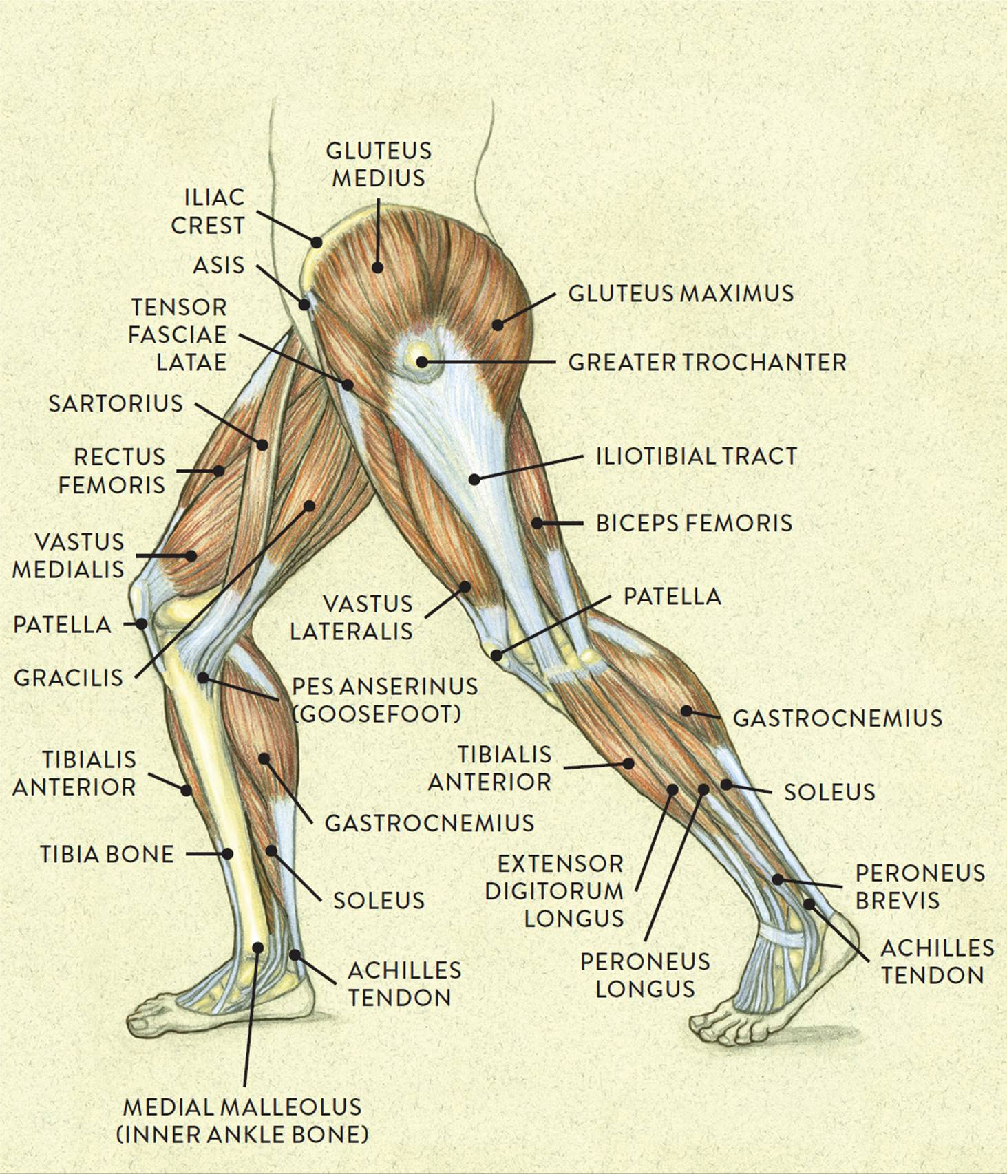
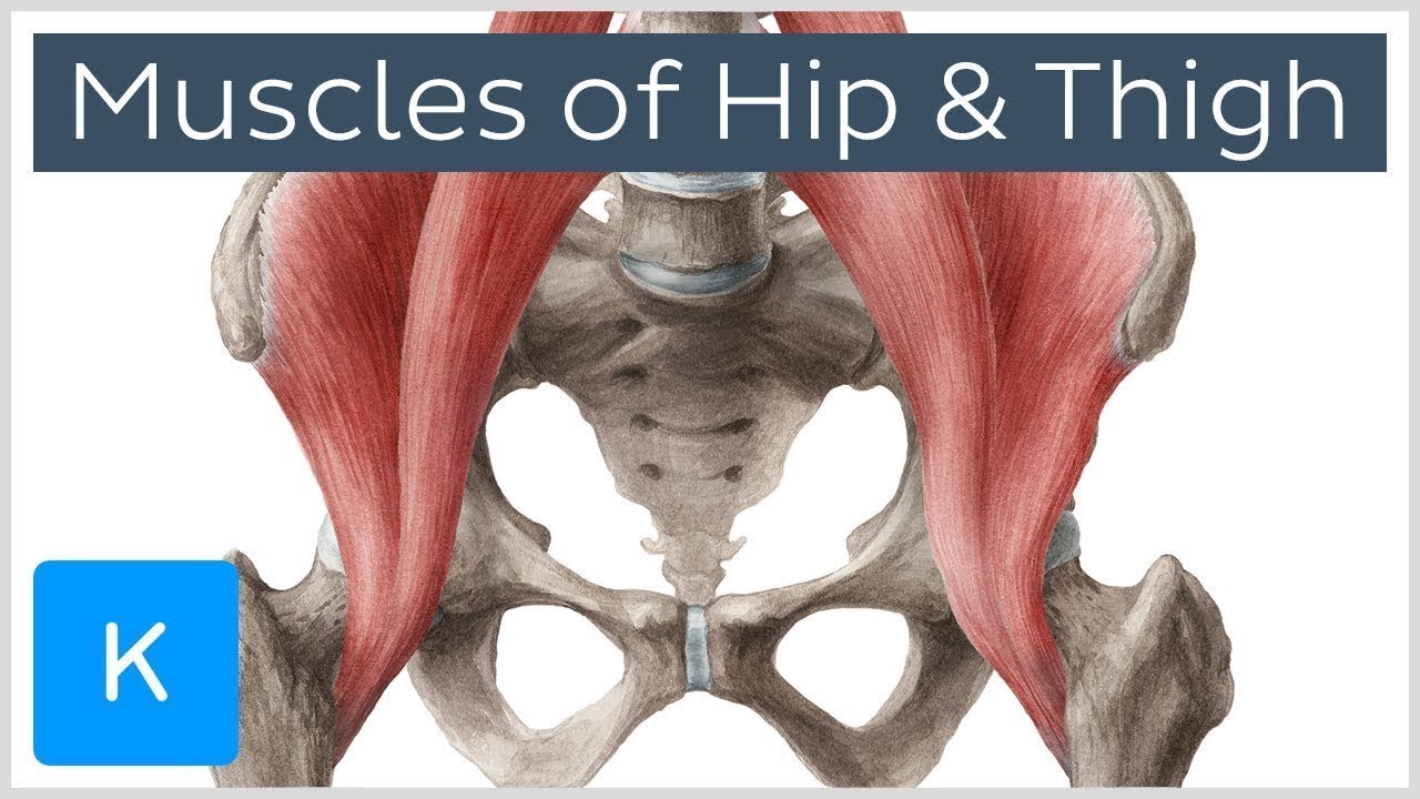
Muscles that move the hip and thigh.
Possibly the most important tendon in terms of mobility is the achilles tendon. The leg muscles are organized in 3 groups: Localized anatomy of the hamstring muscles including semimembranosus, semitendinosus, biceps the hamstrings refer to 3 long posterior leg muscles, the biceps femoris, semitendinosus, and semimembranosus. Human muscles · july 20, 2016. It serves to attach the plantaris, gastrocnemius (calf) and soleus muscles to the calcaneus (heel) bone. We study anatomy at the practical anatomy class we study the human body. The muscle is one of the four quadriceps muscles and is the largest muscle of that group. Lateral (fibular) collateral ligament (fcl) upper part middle part lower part popliteus tendon (pt) upper part i. The posterior upper leg muscles provide your knees with mobility (extension, flexion and rotation) and strength. Muscles that move the hip and thigh. This is the group of muscles that you often see body builders flexing, which protrude just above the knee and take up most of the upper leg. Related posts of muscle anatomy upper leg. Your upper leg includes seven major muscles.
The muscle is one of the four quadriceps muscles and is the largest muscle of that group. This may result in tendon subluxation; Majid doroudi walks you through the the clinical anatomy of the anterior and medial thigh region.produced by dr. Plantarflexes the foot at the ankle joint. Its muscle belly is on the back aspect of the upper arm.
Muscles Of The Hips And Thighs Human Anatomy And Physiology Lab Bsb 141 from s3-us-west-2.amazonaws.com The achilles tendon connects the heel to the calf muscle and is essential for running, jumping. The tendons for these muscles begin at your ischial tuberosity, or ischium (the bony bump under each buttock), and attach on the outer edges of your shinbones (tibia and fibula) just below the back of your knee. The thigh has three sets of strong muscles: A muscle strain (muscle pull or tear) is a common injury, particularly among people who participate in sports. The human leg, in the general word sense, is the entire lower limb of the human body, including the foot, thigh and even the hip or gluteal region. Human muscles · july 20, 2016. Upper leg tendon anatomy : The thigh muscles don't just move your legs.
The human leg, in the general word sense, is the entire lower limb of the human body, including the foot, thigh and even the hip or gluteal region.
Muscle belly splits in two parts. Anterior muscles extend your legs and flex your thighs. The thigh has three sets of strong muscles: It's the area that runs from the hip to the knee in each leg. Related posts of muscle anatomy upper leg. Other muscles of the anterior (front) thigh include the pectineus, sartorius,. Rectus femoris these four muscles come together to form a single tendon, which inserts into the patella, or kneecap. This is why you have to indicate which biceps you are taking about when discussing one or other of these muscles. Plantarflexes the foot at the ankle joint. Muscle anatomy of upper thigh. It is also visible on the medial edge of the thigh from the anterior. This mri wrist coronal cross sectional anatomy tool is absolutely free to use. Lateral (fibular) collateral ligament (fcl) upper part middle part lower part popliteus tendon (pt) upper part i.
Muscle anatomy of upper thigh. This may result in tendon subluxation; Upper leg tendon anatomy from i0.wp.com the achilles tendon or heel cord, also known as the calcaneal tendon, is a tendon at the back of the lower leg, and is the. Ebraheim's educational animated video describes muscle anatomy of the thigh. Tendons are cords made of tough tissue, and they work as special connector pieces between bone and muscle.
Muscles Of The Hip And Thigh Human Anatomy Kenhub Youtube from i.ytimg.com Related online courses on physioplus. On the medial edge of the posterior thigh is the gracilis muscle. They have a lot to do with how your hips move. The leg muscles are organized in 3 groups: It's the area that runs from the hip to the knee in each leg. Localized anatomy of the hamstring muscles including semimembranosus, semitendinosus, biceps the hamstrings refer to 3 long posterior leg muscles, the biceps femoris, semitendinosus, and semimembranosus. Upper leg tendon anatomy : The human leg, in the general word sense, is the entire lower limb of the human body, including the foot, thigh and even the hip or gluteal region.
This mri wrist coronal cross sectional anatomy tool is absolutely free to use.
Muscle belly splits in two parts. Possibly the most important tendon in terms of mobility is the achilles tendon. Upper leg tendon anatomy : It serves to attach the plantaris, gastrocnemius (calf) and soleus muscles to the calcaneus (heel) bone. Upper leg tendon anatomy : Related posts of muscle anatomy upper leg. It also is active in maintaining thigh and kneecap position while walking and. This mri wrist coronal cross sectional anatomy tool is absolutely free to use. Tendons are thick bands of tissue that connect muscles to bone. Upper leg muscle pain is a very hard pain affect the leg pain as a whole. Medial muscles adduct and rotate your thigh, and posterior flex your leg and extend your thigh. •medial thigh muscles•adductor longus muscle•adductor magnus muscle•adductor. Its muscle belly is on the back aspect of the upper arm.

0 Comments:
Posting Komentar